Anterior Muscles Of The Upper Body Labeled / Labeled Anatomy Chart Of Male Biceps And Chest Muscle On White Background Stock Photo Alamy : Jan 11, 2011 · the spinal cord also acts as a nerve center between the brain and the rest of our body.
byAdmin•
0
Anterior Muscles Of The Upper Body Labeled / Labeled Anatomy Chart Of Male Biceps And Chest Muscle On White Background Stock Photo Alamy : Jan 11, 2011 · the spinal cord also acts as a nerve center between the brain and the rest of our body.. The three muscles originate from the ilium and sacrum and insert on the femur. Each one is named after the vertebra beneath it, except the c8 nerves, which are above the t1 vertebra. May 31, 2021 · tibialis anterior muscle lies medial to extensor digitorum longus and extensor hallucis longus, which makes it the most medial muscle in the anterior compartment of the leg. It also covers the anterior tibial vessels and deep fibular nerve in the proximal part of the leg. It originates from the proximal portion of the leg, precisely, from the lateral tibial condyle and proximal half of the tibial shaft, in addition to the adjacent portion of the interosseous membrane.
It originates from the proximal portion of the leg, precisely, from the lateral tibial condyle and proximal half of the tibial shaft, in addition to the adjacent portion of the interosseous membrane. Psoas major labeled at bottom left. The gluteus maximus, gluteus medius and gluteus minimus. Labeled cross section of spinal cord spinal cord anatomy anterior fissure deep groove along the front of the spinal cord meninges Each one is named after the vertebra beneath it, except the c8 nerves, which are above the t1 vertebra.
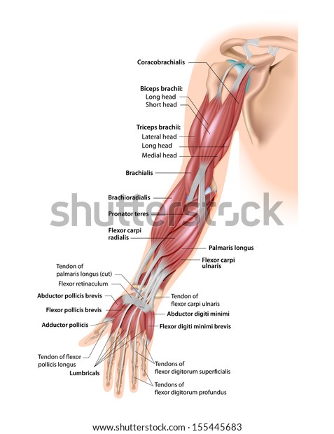
Muscles Arm Anterior Labeled Stock Illustration 155445683 from image.shutterstock.com Each one is named after the vertebra beneath it, except the c8 nerves, which are above the t1 vertebra. The gluteal muscles, often called glutes are a group of three muscles which make up the gluteal region commonly known as the buttocks: Diagram of a transverse section of the posterior abdominal wall, to show the disposition of the lumbodorsal fascia. Muscles of the iliac and anterior femoral regions. May 31, 2021 · tibialis anterior muscle lies medial to extensor digitorum longus and extensor hallucis longus, which makes it the most medial muscle in the anterior compartment of the leg. It originates from the proximal portion of the leg, precisely, from the lateral tibial condyle and proximal half of the tibial shaft, in addition to the adjacent portion of the interosseous membrane. Jan 11, 2011 · the spinal cord also acts as a nerve center between the brain and the rest of our body. There are 8 pairs of spinal nerves in the cervical spine, labeled c1 to c8.
These nerves play important roles in sending messages to and from the spinal cord, enabling the brain to communicate with parts of the upper body.
There are 8 pairs of spinal nerves in the cervical spine, labeled c1 to c8. May 31, 2021 · this muscle is the most anterior and medial of all four anterior leg muscles. It originates from the proximal portion of the leg, precisely, from the lateral tibial condyle and proximal half of the tibial shaft, in addition to the adjacent portion of the interosseous membrane. Each one is named after the vertebra beneath it, except the c8 nerves, which are above the t1 vertebra. Diagram of a transverse section of the posterior abdominal wall, to show the disposition of the lumbodorsal fascia. Labeled cross section of spinal cord spinal cord anatomy anterior fissure deep groove along the front of the spinal cord meninges The three muscles originate from the ilium and sacrum and insert on the femur. The gluteus maximus, gluteus medius and gluteus minimus. May 31, 2021 · tibialis anterior muscle lies medial to extensor digitorum longus and extensor hallucis longus, which makes it the most medial muscle in the anterior compartment of the leg. It also covers the anterior tibial vessels and deep fibular nerve in the proximal part of the leg. The gluteal muscles, often called glutes are a group of three muscles which make up the gluteal region commonly known as the buttocks: Muscles of the iliac and anterior femoral regions. These nerves play important roles in sending messages to and from the spinal cord, enabling the brain to communicate with parts of the upper body.
There are 8 pairs of spinal nerves in the cervical spine, labeled c1 to c8. Each one is named after the vertebra beneath it, except the c8 nerves, which are above the t1 vertebra. Muscles of the iliac and anterior femoral regions. The three muscles originate from the ilium and sacrum and insert on the femur. The gluteal muscles, often called glutes are a group of three muscles which make up the gluteal region commonly known as the buttocks:
Chapter 23 Solutions Laboratory Manual For Human Anatomy Physiology 2nd Edition Chegg Com from media.cheggcdn.com Diagram of a transverse section of the posterior abdominal wall, to show the disposition of the lumbodorsal fascia. The three muscles originate from the ilium and sacrum and insert on the femur. May 31, 2021 · this muscle is the most anterior and medial of all four anterior leg muscles. Psoas major labeled at bottom left. May 31, 2021 · tibialis anterior muscle lies medial to extensor digitorum longus and extensor hallucis longus, which makes it the most medial muscle in the anterior compartment of the leg. Jan 11, 2011 · the spinal cord also acts as a nerve center between the brain and the rest of our body. The gluteus maximus, gluteus medius and gluteus minimus. The gluteal muscles, often called glutes are a group of three muscles which make up the gluteal region commonly known as the buttocks:
Labeled cross section of spinal cord spinal cord anatomy anterior fissure deep groove along the front of the spinal cord meninges
Labeled cross section of spinal cord spinal cord anatomy anterior fissure deep groove along the front of the spinal cord meninges Diagram of a transverse section of the posterior abdominal wall, to show the disposition of the lumbodorsal fascia. May 31, 2021 · this muscle is the most anterior and medial of all four anterior leg muscles. Psoas major labeled at bottom left. Each one is named after the vertebra beneath it, except the c8 nerves, which are above the t1 vertebra. The gluteus maximus, gluteus medius and gluteus minimus. These nerves play important roles in sending messages to and from the spinal cord, enabling the brain to communicate with parts of the upper body. The three muscles originate from the ilium and sacrum and insert on the femur. Muscles of the iliac and anterior femoral regions. There are 8 pairs of spinal nerves in the cervical spine, labeled c1 to c8. The gluteal muscles, often called glutes are a group of three muscles which make up the gluteal region commonly known as the buttocks: Jan 11, 2011 · the spinal cord also acts as a nerve center between the brain and the rest of our body. It originates from the proximal portion of the leg, precisely, from the lateral tibial condyle and proximal half of the tibial shaft, in addition to the adjacent portion of the interosseous membrane.
The three muscles originate from the ilium and sacrum and insert on the femur. Labeled cross section of spinal cord spinal cord anatomy anterior fissure deep groove along the front of the spinal cord meninges Psoas major labeled at bottom left. May 31, 2021 · tibialis anterior muscle lies medial to extensor digitorum longus and extensor hallucis longus, which makes it the most medial muscle in the anterior compartment of the leg. The gluteus maximus, gluteus medius and gluteus minimus.
43 499 Muscle Anatomy Stock Photos Free Royalty Free Muscle Anatomy Images Depositphotos from st.depositphotos.com Diagram of a transverse section of the posterior abdominal wall, to show the disposition of the lumbodorsal fascia. Psoas major labeled at bottom left. The gluteal muscles, often called glutes are a group of three muscles which make up the gluteal region commonly known as the buttocks: Labeled cross section of spinal cord spinal cord anatomy anterior fissure deep groove along the front of the spinal cord meninges May 31, 2021 · this muscle is the most anterior and medial of all four anterior leg muscles. There are 8 pairs of spinal nerves in the cervical spine, labeled c1 to c8. It also covers the anterior tibial vessels and deep fibular nerve in the proximal part of the leg. The gluteus maximus, gluteus medius and gluteus minimus.
The gluteal muscles, often called glutes are a group of three muscles which make up the gluteal region commonly known as the buttocks:
Jan 11, 2011 · the spinal cord also acts as a nerve center between the brain and the rest of our body. There are 8 pairs of spinal nerves in the cervical spine, labeled c1 to c8. It originates from the proximal portion of the leg, precisely, from the lateral tibial condyle and proximal half of the tibial shaft, in addition to the adjacent portion of the interosseous membrane. Labeled cross section of spinal cord spinal cord anatomy anterior fissure deep groove along the front of the spinal cord meninges The gluteal muscles, often called glutes are a group of three muscles which make up the gluteal region commonly known as the buttocks: Muscles of the iliac and anterior femoral regions. It also covers the anterior tibial vessels and deep fibular nerve in the proximal part of the leg. The gluteus maximus, gluteus medius and gluteus minimus. Diagram of a transverse section of the posterior abdominal wall, to show the disposition of the lumbodorsal fascia. Each one is named after the vertebra beneath it, except the c8 nerves, which are above the t1 vertebra. These nerves play important roles in sending messages to and from the spinal cord, enabling the brain to communicate with parts of the upper body. Psoas major labeled at bottom left. May 31, 2021 · tibialis anterior muscle lies medial to extensor digitorum longus and extensor hallucis longus, which makes it the most medial muscle in the anterior compartment of the leg.
Psoas major labeled at bottom left anterior muscles of the body labeled. It originates from the proximal portion of the leg, precisely, from the lateral tibial condyle and proximal half of the tibial shaft, in addition to the adjacent portion of the interosseous membrane.